- 진단 장비
- 광간섭 단층촬영
DRI OCT Triton series
TOPCON의 Swept Source OCT Angio™는 OCT Angiography와 Swept Source OCT 기술 그리고 1,050nm의 장 파장 기술을 결합한 제품입니다. 독자적인 영상 처리 알고리즘인 OCTARA™을 사용하여 고감도의 혈관조영 검출 기능을 제공해 혈관의 구조를 시각화할 수 있습니다.

Key Features
 고밀도의 Swept Source OCT
 MediaOpacity를 침투 하는 깊은 투과력
 멀티 모달 이미징
 Swept Source OCT Angiography
 녹내장 진료를 위한 Hood Report
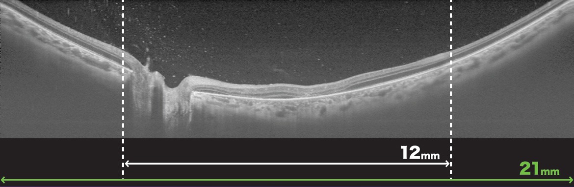
 Wide-Field OCT 최대 21mm Scan
 Wide-Field OCT Angiography
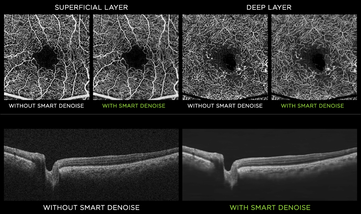
 Smart Denoise
Courtesy: Dr. Yoshinori Mitamura, MD, PhD, Tokushima University, Japan
광범위한 OCT와 OCT-A 이미지를 통해 더 다양한 임상 인사이트를 얻을 수 있습니다. 이는 다양한 케이스에 활용할 수 있습니다.
Courtesy: Prof. Christopher Leung, The University of Hong Kong.
Topcon의 독자적인 알고리즘을 통해, 조밀한 데이터 큐브 내의 모든 B-스캔에서 노이즈가 감소된 고품질의 OCT 및 OCT-A 영상이 생성됩니다.
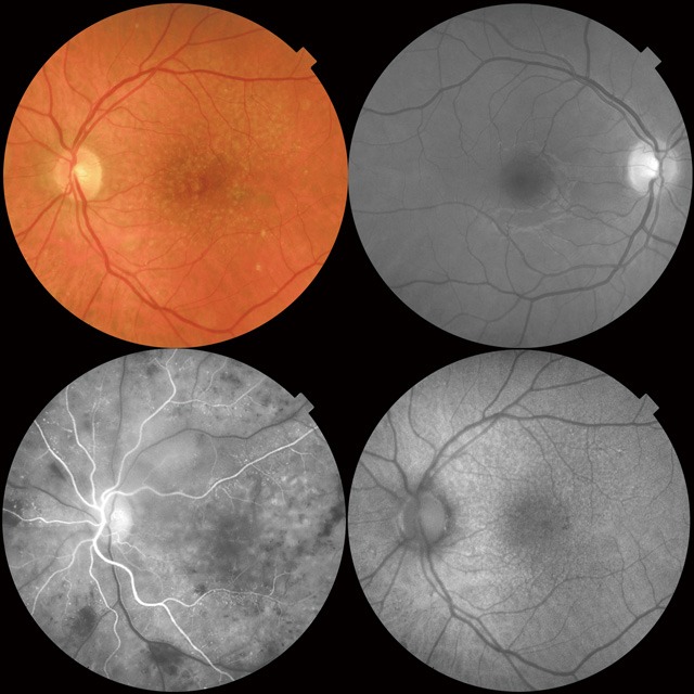
형광안저촬영(FA) 및 자가형광영상(FAF) 기능은 Triton Plus의 진단 능력을 더욱 향상시킵니다.
이 All-in-one 장비는 효율적인 임상 워크플로우를 가능케 합니다.
DRI OCT Triton은 OCT 이미지과 안저 이미지를 한 번의 촬영으로 동시에 획득할 수 있습니다.
PinPoint™ 기능은 안저 영상 내에서 B-Scan 위치를 식별할 수 있게 해줍니다.
B-Scan과 안저 영상을 비교함으로써 보다 효율적인 임상 진단이 가능합니다.
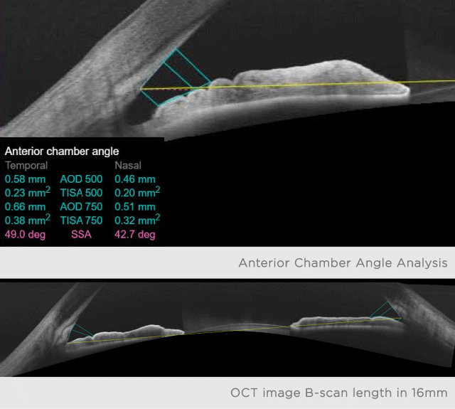
전안부 렌즈는 정량적 분석 기능도 함께 제공됩니다.
새롭게 업데이트된 전안부 기능은 종합적인 안과 진료 환경에서 Triton을 효율적으로 활용할 수 있게 합니다.
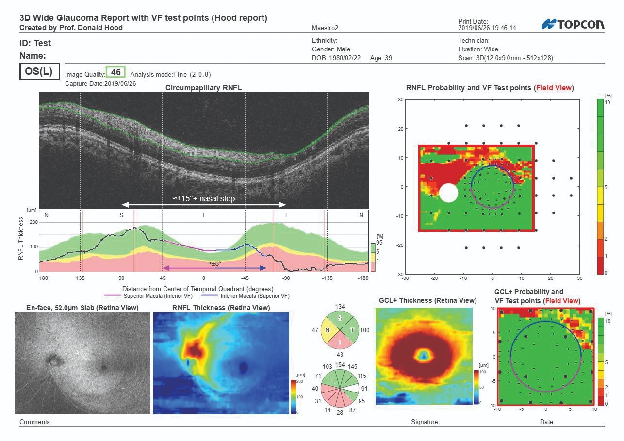
Hood Report는 망막 두께, RNFL(망막신경섬유층), GCL(신경절세포층), 그리고 시신경 주변 지표를 한 번의 스캔으로 제공합니다.
이 리포트는 구조적 확률 맵(GCC/RN리)과 기능적 정보(시야검사 위치 오버레이)를 연계함으로써 의사결정 과정을 줄어듭니다.
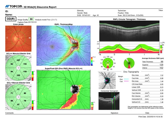
12×9mm Wide Scan은 시신경과 황반을 모두 포함하며, 한 번의 촬영으로 Posterior pole 전체에 대한 종합적인 평가를 제공합니다.
또한, 레퍼런스 데이터베이스와의 비교 분석도 가능합니다.
Note: 이 웹사이트에 명시되어 있는 정보는 의료 전문가를 위한 것입니다. 모든 제품, 서비스, 제안이 모든 시장에서 승인 또는 제공되는 것이 아니며 판매되는 제품이 국가마다 다를 수 있습니다.국가별 정보 및 구매 가능 제품에 대한 내용은 지역 배급사에 문의하십시오.
상세 정보 요청하기
제품에 대한 문의사항이 있으시거나 구매 의사가 있으신 경우 등 언제든지 도움을 드리고자 합니다.
아래 양식을 작성해 주시면 연락을 드리도록 하겠습니다.


